Miklossy J, Kraftsik R, Pillevuit O, Lepori D, Genton C, Bosman FT. Neuropathol Exp Neurol. 1998 Dec;57(12):1202-12.Curly fiber and tangle-like inclusions in the ependyma and choroid plexus--a pathogenetic relationship with the cortical Alzheimer-type changes?
Abstract
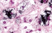
The question of whether thread- and tangle-like inclusions of the choroid plexus (known as Biondi inclusions) are related to the cortical lesions in Alzheimer disease (AD) has been debated for almost a century, yet remains unanswered. Recently beta-amyloid protein was biochemically isolated from the plexus, indicating a possible pathogenetic relationship between the degenerative changes of the cerebral cortex and those of the plexus. The goal of the present study was to analyze whether or not a significant correlation exists between the occurrence of the cortical AD-type changes and those in the ependyma and choroid plexus. In 292 consecutive autopsy cases several cortical areas, the ependyma, and the choroid plexus were analyzed to look for AD-type changes and Biondi inclusions using histochemical staining techniques and immunohistochemistry. A semiquantitative analysis of the density of cortical AD-type changes showed that of the 292 cases, 63 had severe cortical changes, 23 moderate changes, and 142 discrete changes. In 64 cases no plaques or neurofibrillary tangles were found. The number of cases with thread- and tangle-like elements in the plexus and ependyma was more than 96% in the 3 groups with cortical AD-type lesions, but low in the group without AD-type cortical changes (19%). The pathological argyrophilic filaments accumulating in the ependymal layer and plexus had histochemical properties of amyloid and were immunoreactive with antibodies to P component, ubiquitin, fibronectin and Tau protein. They did not react with antibodies to neurofilament proteins. Ultrastructurally, they consisted of densely packed straight and paired helical filaments and closely resembled neurofibrillary tangles and neuropil threads. The highly significant correlation (chi2, p = 0.001; R = 0.85) between the occurrence of AD-type changes in the cortex and those in ependyma and plexus suggests a pathogenetic Relationship.
Miklossy J, Taddei K, Martins R, Escher G, Kraftsik R, Pillevuit O, Lepori D, Campiche M. J Neuropathol Exp Neurol. 1999 Aug;58(8):803-14.Alzheimer disease: curly fibers and tangles in organs other than brain.
Abstract
The filamentous brain lesions that define Alzheimer disease (AD) consist of senile plaques and neurofibrillary tangles. Undulated pathological filaments--curly fibers or neuropil threads--also occur in the neuropil. Beta-amyloid precursor proteins are synthesized by many cells outside the central nervous system and recently, deposition of beta-amyloid-protein was reported to occur in non-neuronal tissues. In addition, increasing data claim the importance of chronic inflammation in the pathogenesis of AD. These observations suggest that AD may be a widespread systemic disorder. Here we report that pathological argyrophilic filaments with histochemical properties of amyloid showing striking morphological similarity to curly fibers and/or tangles accumulate not only in ependymal layer and in epithelial cells of choroid plexus, but also in several other organs (e.g. liver, pancreas, ovary, testis, thyroid) in AD. The ependyma, choroid plexus, and various organs of 39 autopsy cases were analyzed. In search of curly fiber and tangle-like changes in organs other than brain, 395 blocks from 21 different tissues of 24 AD cases, 5 cases with discrete or moderate AD-type changes, and 10 control cases were investigated. We found in non-neuronal cells "curly fibers" or "tangles" immunoreactive with antibodies to P component, Tau-protein, ubiquitin, fibronectin, and Apolipoprotein-E, but lacking immunoreactivity with antibodies to neurofilament proteins. Ultrastructurally they consist of densely packed straight and paired helical filaments and closely resemble neurofibrillary tangles and neuropil threads. These observations indicate that the formation of "curly fibers" and "tangles" is not unique to the central nervous system. The results suggest that AD might be a systemic disorder or that similar fibrillary changes to tangles and curly fibers may also be associated with other amyloidosis than beta-amyloidosis. Further investigations are necessary to understand the pathogenetic interest of these fibrillary changes outside the CNS.